Arteries Diagram / Coronary Arteries And Heart Disease. There is a point at which the anterior and posterior arterial circuits of the brain unite or anastomose. Learn the differences between an artery and a vein. The veins also lack the elastic internal lamina that lies. Most arteries carry oxygenated blood; The two exceptions are the pulmonary and the umbilical arteries, which carry deoxygenated blood to the organs that oxygenate it (lungs and placenta.
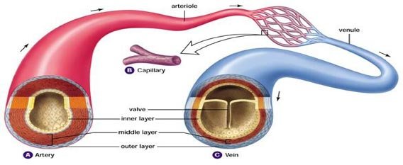
The tunica medica, which is the very muscular middle layer in arteries, is thinner and less muscular in veins. This area is known as the circle of willis. Because the rest of the body, and most especially the brain, needs a steady supply of oxygenated blood that is free of all but the slightest. Systemic arteries deliver blood to the rest of the body. It originates from the heart and branches out into smaller arteries which supply blood to the head region (brachiocephalic artery), the heart itself (coronary arteries), and the lower regions of the body.
Arteries And Their Function Diagram Quizlet from o.quizlet.com These arteries arise in the neck, and ascend to the cranium. These vessels are channels that distribute blood to the body. The vertebral arteries, and the internal carotid arteries. The coronary arteries wrap around the outside of the heart. Anatomynote.com found blood circulation principal veins and arteries diagram from plenty of anatomical pictures on the internet. An artery (plural arteries) (from greek ἀρτηρία (artēria) 'windpipe, artery') is a blood vessel that takes blood away from the heart to one or more parts of the body (tissues, lungs, brain etc.). This is the area where speech, thinking, sensory, motor, and personality functions reside. We hope this picture human body artery diagram in detail can help you study and research.
Clogged lumbar arteries are far more common than most people realize.
After receiving blood directly from the left ventricle of the heart, the. This allows blood to flow around the blocked artery to another artery nearby or to the same artery past the blockage, protecting the heart tissue from injury. Fatty deposits accumulated in carotid arteries can make the arteries get narrowed, thus decreasing blood flow to your brain or make them completely blocked. Original vintage human anatomy victorian bookplate print 1890s medical diagram veins arteries blood circulatory system of the human body thepapermuseum. For more anatomy content please follow us and visit our website: The typical configuration consists of two coronary arteries, a left main coronary artery (lmca) and a right coronary artery (rca), arising from the left posterior and right anterior aortic or coronary sinuses respectively, in the proximal ascending aorta.these are the only two branches of the ascending aorta. Spontaneous coronary artery dissection — sometimes referred to as scad — is an uncommon emergency condition that occurs when a tear forms in a blood vessel in the heart. Coronary arteries supply blood to the heart muscle. Arteries of the brain and 'circle of willis' diagram. The vertebral arteries, and the internal carotid arteries. This is the area where speech, thinking, sensory, motor, and personality functions reside. The narrowed arteries are at higher risk for complete blockage from a sudden. We hope this picture blood circulation principal veins and arteries diagram can help you study and research.
This allows blood to flow around the blocked artery to another artery nearby or to the same artery past the blockage, protecting the heart tissue from injury. The right coronary artery courses in the right atrioventricular groove. Ascending aorta, aortic arch, thoracic aorta, and abdominal aorta. Anatomynote.com found blood circulation principal veins and arteries diagram from plenty of anatomical pictures on the internet. Original vintage human anatomy victorian bookplate print 1890s medical diagram veins arteries blood circulatory system of the human body thepapermuseum.
Difference Between Arteries And Veins Table Easy Biology Class from www.easybiologyclass.com Coronary circulation is the circulation of blood in the blood vessels that supply the heart muscle (myocardium). Coronary arteries supply oxygenated blood to the heart muscle, and cardiac veins drain away the blood once it has been deoxygenated. We hope this picture blood circulation principal veins and arteries diagram can help you study and research. Though more often occurring with carotid arteries (the other major ones supplying the brain through the neck), vertebral arteries can be impacted. This is the area where speech, thinking, sensory, motor, and personality functions reside. Blood is pumped from the heart in the arteries. Transposition of the great arteries is usually detected either prenatally or within the first hours to weeks of life. Each of these arteries delivers blood to the leg and continues into the foot, with the posterior tibial and fibular arteries forming the plantar arteries and plantar arch that supply blood to the bottom of the foot and toes.
This allows blood to flow around the blocked artery to another artery nearby or to the same artery past the blockage, protecting the heart tissue from injury.
Arteries carry blood away from the heart in two distinct pathways: The typical configuration consists of two coronary arteries, a left main coronary artery (lmca) and a right coronary artery (rca), arising from the left posterior and right anterior aortic or coronary sinuses respectively, in the proximal ascending aorta.these are the only two branches of the ascending aorta. In fact, about 10 percent of americans have already begun to form blockages in lumbar arteries by the time they're 20. Corrective surgery soon after birth is the usual treatment for transposition of the great arteries. When the coronary arteries narrow to the point that blood flow to the heart muscle is limited (coronary artery disease), collateral vessels may enlarge and become active. Blood is pumped from the heart in the arteries. Learn vocabulary, terms, and more with flashcards, games, and other study tools. We think this is the most useful anatomy picture that you need. These arteries arise in the neck, and ascend to the cranium. The veins also lack the elastic internal lamina that lies. Classifica'on*of*arteries* • elas'c*arteries* *(conduc'ng*arteries) *aorta,*brachiocephalic,* commoncarod,* subclavian, vertebral,pulmonary,common* iliac. Original vintage human anatomy victorian bookplate print 1890s medical diagram veins arteries blood circulatory system of the human body thepapermuseum. Coronary circulation is the circulation of blood in the blood vessels that supply the heart muscle (myocardium).
Circle of willis is indeed a hot neuroanatomy topic! Blood is transported in arteries, veins and capillaries. 5 out of 5 stars (293) 293 reviews $ 24.27. This area is known as the circle of willis. Because the rest of the body, and most especially the brain, needs a steady supply of oxygenated blood that is free of all but the slightest.
The Arteries Human Anatomy Picture Definition Conditions More from img.webmd.com Circle of willis is indeed a hot neuroanatomy topic! Coronary arteries supply blood to the heart muscle. We hope this picture blood circulation principal veins and arteries diagram can help you study and research. It is a central communication that unites the internal carotid and vertebrobasilar systems. The right coronary artery courses in the right atrioventricular groove. The narrowed arteries are at higher risk for complete blockage from a sudden. It is returned to the heart in the veins. Ascending aorta, aortic arch, thoracic aorta, and abdominal aorta.
Anatomynote.com found human body artery diagram in detail from plenty of anatomical pictures on the internet.
These vessels are channels that distribute blood to the body. After receiving blood directly from the left ventricle of the heart, the. The two exceptions are the pulmonary and the umbilical arteries, which carry deoxygenated blood to the organs that oxygenate it (lungs and placenta. Original vintage human anatomy victorian bookplate print 1890s medical diagram veins arteries blood circulatory system of the human body thepapermuseum. We hope this picture human body artery diagram in detail can help you study and research. There is a point at which the anterior and posterior arterial circuits of the brain unite or anastomose. This area is known as the circle of willis. We think this is the. Spontaneous coronary artery dissection — sometimes referred to as scad — is an uncommon emergency condition that occurs when a tear forms in a blood vessel in the heart. The coronary arteries wrap around the outside of the heart. Running behind the duct that allows urine to flow from the kidneys to the bladder (ureter) in its upper portion, this artery courses down the body with its corresponding vein in front of it.the artery branches at the rear (posterior) and front of the body and supplies blood to various muscle groups, bones, nerves, and organs in and around the pelvis. Ascending aorta, aortic arch, thoracic aorta, and abdominal aorta. The anterior tibial artery forms the arcuate artery and its many branches to supply blood to the top of the foot.
Share :
Post a Comment
for "Arteries Diagram / Coronary Arteries And Heart Disease"


Post a Comment for "Arteries Diagram / Coronary Arteries And Heart Disease"